
Geometric model with a 7.5 mm bulb and a 4.5 mm distal internal carotid artery (ratio 1.67, the same one implicit in the Rothwell line). The blue dashed lines extend the distal ICA calibre into the bulb: the plaque starts to count for NASCET only when the residual channel crosses them. With a wider or narrower bulb the same plaque would give different numbers — which is why the conversion between the two methods is not a law of nature.
Measured diameters
In millimetres, from B-mode, CT angiography or MR angiography. A and C are enough for NASCET alone, A and B for ECST alone.
NASCET
—
Enter A and C
ECST
—
Enter A and B
Expected ICA PSV
—
SRU 2003 consensus
How to measure
Where A, B and C are measured
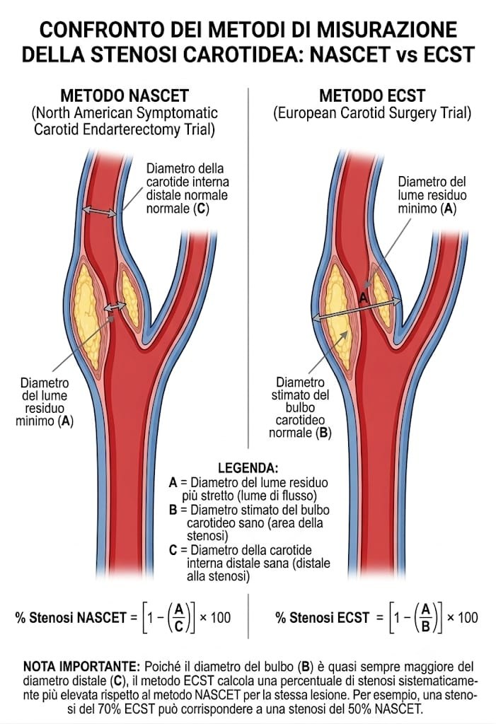
Measuring C too close to the plaque, where the vessel is still tapering, inflates the NASCET value; measuring it on a hypoplastic or low-flow internal carotid lowers it. B is not the outer diameter of the vessel including the wall, but the lumen calibre the bulb would have had without disease. When calcification with acoustic shadowing is present, the B-mode lumen measurement must always be compared with the velocity data.
Interpretation
Why the same plaque gives two different numbers
NASCET uses as its denominator the healthy internal carotid beyond the plaque, a vessel smaller than the bulb: the smaller denominator lowers the percentage. ECST estimates the calibre of the bulb as it was before the disease, a larger denominator, and therefore gives higher values for the same lesion. The difference is not a measurement error: they are two different rulers laid on the same vessel.
The main guidelines on indications for treatment (ESVS, AHA/ASA, SVS) and the duplex velocity criteria built into the carotid form of VASCULAR REPORT are expressed in the NASCET convention. If an external report or an angiogram gives an ECST value, it must be converted before those thresholds are applied.
| Grade | NASCET stenosis | Equivalent ECST |
|---|---|---|
| Mild | < 50% | < 70% |
| Moderate | 50 – 69% | 70 – 81% |
| Severe | 70 – 99% | 82 – 99% |
| Near-occlusion | “string sign”, collapsed distal ICA | — |
| Occlusion | 100% (A = 0, no residual lumen) | 100% |
The ECST column applies only from NASCET 30% (= ECST 58%) upwards: below that threshold the equivalence line cannot be applied (see below).
The Rothwell line ECST = 0.6 × NASCET + 40 amounts, in geometric terms, to assuming a fixed ratio between the bulb and the distal internal carotid: the coefficient 0.6 is the C/B ratio, i.e. a bulb about 1.67 times as wide as the downstream vessel. The 40-point intercept follows from this assumption, and with it the paradoxical behaviour at low grades: the formula assigns ECST 40% to a carotid that NASCET reads as 0%, ECST 50% to a NASCET of 17%, and negative values below ECST 40%. This is not a calculation error but the limit of a regression derived from symptomatic stenoses and extrapolated outside its range. For this reason the calculator does not convert below NASCET 30% (= ECST 58%): at those grades the plaque is described and the velocity parameters are reported, rather than translating one percentage into another.
With plaque confined to the bulb and a healthy distal internal carotid, NASCET can give very low percentages compared with the severity seen on the image, down to negative values when the residual lumen exceeds the distal calibre. In these cases B-mode and velocities remain the primary reference, as provided by the IAC 2023 criteria used in the carotid form. In near-occlusion, by contrast, collapse of the downstream internal carotid reduces C and the NASCET percentage underestimates severity: near-occlusion must be recognised and described, not calculated.
What to write in the report
- Always state the method next to the percentage: “70% stenosis by NASCET”, not “70% stenosis”.
- If the grade comes from a converted ECST value, say so: a conversion does not carry the same weight as a direct measurement.
- Report the velocity parameters (ICA PSV and EDV, ICA/CCA ratio) alongside the morphological data: in duplex scanning they remain the main grading criterion.
References
Sources
- NASCET Collaborators (N Engl J Med 1991;325:445–53) — Beneficial effect of carotid endarterectomy in symptomatic patients with high-grade carotid stenosis.
- European Carotid Surgery Trialists' Collaborative Group (Lancet 1998;351:1379–87) — Randomised trial of endarterectomy for recently symptomatic carotid stenosis: final results of the MRC European Carotid Surgery Trial.
- Rothwell PM et al. (Stroke 1994;25:2435–9) — Equivalence of measurements of carotid stenosis: a comparison of three methods on 1001 angiograms.
- Grant EG et al. (Radiology 2003;229:340–6) — Carotid artery stenosis: gray-scale and Doppler US diagnosis, Society of Radiologists in Ultrasound consensus conference.
- ESVS 2023 — Clinical Practice Guidelines on the Management of Atherosclerotic Carotid and Vertebral Artery Disease.
Quick-reference tool for healthcare professionals. It does not replace the original guidelines, direct remeasurement on the image or the specialist's clinical judgement. All calculations run in the browser: no patient data is transmitted to or stored on any server.